
A microscope is an instrument used to see objects that are too small to be seen by the naked eye. It comprised from several lenses through which the light passes through, destined to focus the image and lessening the blur seen through the eye piece.
The optical microscope is divided into three main parts:
The microscope head, being set on top, which holds the optical parts; The microscope base, supporting the microscope and from which the light reflecting on the lens emerges and goes through the sample; and third, the supporting arm connecting between the head and base.
Microscopes come in different configuration according to the eye pieces – Monocular, Binocular and Trinocular.
Monocular comes from joining “mono” – alone, and “Ocular” – eye; from here it’s easy to deduce the monocular has only one eyepiece. It was the first instrument used to enlarge objects with limited field depth. Today it is rare to encounter the monocular outside of a museum.
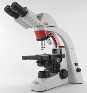
The binocular, as expected, is a microscope with two eyepieces in its head, and is very common in every lab. The binoculars come in various models and qualities, from simple and efficient models such as BA210RED or from the RED220 series and up to the top of the line smart microscopes such as Motic’s Panthera-L model, which comes with built-in PC, camera and Wi-Fi broadcasting.
![]()
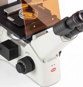
The trinocular is very similar to the binocular as for its properties, but with two standard eyepieces and one camera eyepiece in which an external camera is placed. In trinocular microscope sometimes the light is reflected towards the eyepieces or towards the camera, and in other models it is possible to watch both the eyepieces and the camera simultaneously. An excellent example for quality trinocular models is the RED223 model from Motic’s excellent series RED220 or the Panthera-U model.
In general, all optic microscopes (either binocular or trinocular) are used to watch slides carrying thin samples, through which the light can pass. When using 3D samples we recommend working with either a stereoscope or the digital microscopes from Dino-Lite.
But what is a stereoscope? The main difference between it and a regular microscope is the field depth seen via the eyepieces; it contains upright illumination in addition to the back light, making it possible to examine non-transparent samples. Its goal is to create a 3D image (stereoscopic) similar to the way we see. Each one of the eyepieces displays a slightly different image, which interprets in the brain to a single, combined, 3D image. An example for a stereoscope is the excellent SMZ-161 series by Motic. And of course, the stereoscope could be binocular or trinocular. As discussed before, we will use a stereoscope when looking at samples with volume such as organs or tissue samples (plants/animals).
Another type of microscope is the inverted microscope, where the light source us from above while the objectives come from below – thus, inverted. It was designed to watch cell cultures at the bottom of flasks or wells, as they live a liquid medium.
The most recent developments in light microscope largely center on the rise of fluorescence microscopy in biology. During the last decades of the 20th century, particularly in the post-genomic era, many techniques for fluorescent staining of cellular structures were developed. The main groups of techniques involve targeted chemical staining of particular cell structures, for example, the chemical compound DAPI to label DNA which is being used to identify and count cells nuclei.
An excellent example for an inverted microscope (with fluorescent filters option) is AE31 by Motic.
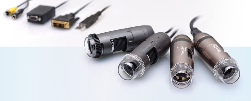
In addition to the standard optical microscope you are also able to find mobile, digital hand-held microscopes by Dino-Lite. Those professional microscopes have similar properties to the optical stereoscope but are much smaller (as a pocket flashlight). The Dino-Lite are enlarging and documenting microscopes and are intended to display the image on a screen – your PC or Android.
The digital microscope is comprised from built-in LED lights as an upright illumination. It has full magnification range, manual focus and is able to connect to a PC, tablet or Android and show the image with no eyepiece, while documenting and capturing images and videos. There are over 150 models varying in wave length (white, UV, fluorescent), maximal magnification (x220, x470, 700-900x), working distance, resolution and many more customizable features.
Usually you could find the Dino-Lite microscopes in use at labs and fields to examine 3D samples with documenting images and videos.
Keep in mind that most people tend to mix magnification and resolution. Magnification looks at the size of the examined object and resolution looks at number of pixels and thus directly effecting the picture quality.
In conclusion, there are many types of microscopes and matching the correct model to your specific application is crucial to the examination success and correct documentation. We here at Iner-Tech believe you have to match the correct instrument and its features to your needs, and so we come and personally demonstrate the various models at your preferred location, making sure it fits prior to the customer purchasing it.
Having trouble choosing the correct conformation? Contact us today to set a personal DEMO meeting!